Oct Retinal Nerve Fiber Layer
Optical coherence tomography of the optic disc Dramatic visual recovery in untreated indirect traumatic optic neuropathy Layer fiber retinal nerve thickness optical coherence foto tomography disc optic imaging using gr
Dramatic Visual Recovery in Untreated Indirect Traumatic Optic Neuropathy
Layer oct retinal glaucoma fiber volume nerve detection dimensional measurement three Retinal nerve thickness rnfl macular scans spectralis macula etdrs Nerve retinal rnfl rpe interpretation pigment
Retinal photography & optical coherence tomography: prominent retinal
Journals nerve retinal layer fiber glaucoma large myopiaHeidelberg nerve spectralis retinal fiber rnfl engineering damage glaucomatous Optic oct nerve cirrus thinning imaging retinal neuropathy layer fiber ganglion traumatic cell visual os pallor indicating cases dramatic recoveryRetinal nerve fiber layer analysis from spectralis-oct (heidelberg.
Nerve layer fiber retinal prominent photographyNerve oct optic quadrants fiber layer atrophy papillitis vaccine induced suspected term teaching follow case report long retinal showed thinning Myelinated nerve fiber layer defect nfl myelin raphe horizontal rnfl visual fieldNotch defect oct eye nerve optic glaucoma disc layer fiber retinal nfl angle open tomography optical coherence poag primary test.

Three-dimensional retinal nerve fiber layer volume measurement with oct
Retinal nerve fiber layer imaging in myopiaVisual nerve fiber layer retinal fields defects figure loss seen oct spatially deep well Representative spectralis sd-oct scans of (a) retinal nerve fiber layerWoman experiences decreased vision with normal fundus exam.
Flashes floaters referred blurry vision woman light oct eye macula left figure rightFigure 1 from deep defects seen on visual fields spatially correspond Atlas entryInterpretation of oct scan. rnfl, retinal nerve fiber layer; rpe.

Normal fundus experiences decreased exam vision woman
Woman referred for blurry vision, flashes of light and floatersAtlas entry Long-term follow-up of suspected vaccine-induced papillitis: a teachingMyelinated retinal nerve fiber layer.
Myelinated nerve fiber retinal layer fundus rnfl eyewiki mild yo hyperopia vision girl .


Representative Spectralis SD-OCT scans of (A) retinal nerve fiber layer

Three-dimensional Retinal Nerve Fiber Layer Volume Measurement with OCT

Atlas Entry - Optic Disc Notch and Retinal Nerve Fiber Layer Defect in

Woman experiences decreased vision with normal fundus exam
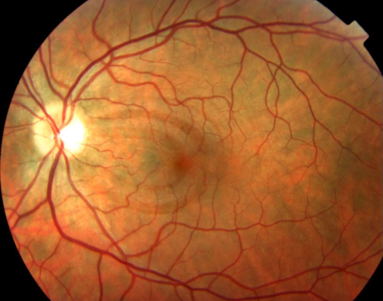
Retinal Photography & Optical Coherence Tomography: Prominent Retinal

Myelinated Retinal Nerve Fiber Layer - EyeWiki

Optical Coherence Tomography of The Optic Disc | EYE DAY CLINIC

Long-term Follow-up of Suspected Vaccine-Induced Papillitis: A Teaching

Interpretation of OCT scan. RNFL, retinal nerve fiber layer; RPE